The Role of Digital Pathology in Solving Specialized Tissue Diagnosis Challenges
Diagnosis of Complex Neoplasms
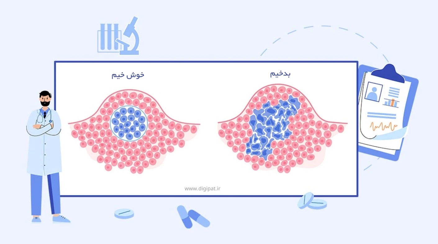
One of the major challenges in traditional pathology is the accurate identification and diagnosis of poorly differentiated neoplasms. These neoplasms are often difficult to distinguish from inflammatory lesions or non-neoplastic lesions due to their high similarity. Pathologists typically spend a significant amount of time examining very subtle changes in cells and tissues, which can lead to diagnostic errors. Additionally, in cases where the tissue has undergone necrosis or severe damage, identifying specific cellular features becomes even more challenging.
Treatment Prediction with Tissue Markers
Biomarkers such as HER2, ER, and PR play a key role in determining the treatment pathway for many patients. However, the immunohistochemical analysis of these markers in traditional pathology heavily relies on the visual interpretation of the pathologist. This leads to inaccuracies and lack of reproducibility in results across different laboratories. Even when experienced pathologists are involved, differences in staining methods and sample preparation can affect the results.
Shortage of Specialists
With the growing population and increasing number of patients, the demand for pathology services has also risen. In many regions, the shortage of skilled pathologists has become a crisis. This high workload can reduce diagnostic accuracy, as pathologists are forced to examine a large number of slides in a short time. This issue also causes patients to wait longer for their diagnostic results.
Analysis of Rare Tissues
Rare tissues or cases with highly unusual changes require detailed examination and sometimes consultation with multiple specialists. In traditional pathology, sending samples to specialized laboratories or renowned experts can take a long time, and there is a risk of sample damage during transportation. Additionally, limited access to similar data for comparison and research is another obstacle.

Solutions Offered by Digital Pathology
Artificial Intelligence in Image Analysis
Artificial intelligence and deep learning in digital pathology image analysis have the ability to identify specific microscopic patterns that may even be overlooked by experienced pathologists. Advanced algorithms can accurately identify neoplasms based on specific features such as cell shape, nucleus size, and tissue density. This technology not only increases diagnostic accuracy but also significantly speeds up the examination of samples.
Quantitative Immunohistochemical Analysis
One of the significant achievements of digital pathology is the ability to quantitatively analyze biomarker expression. Unlike traditional methods, which are limited to qualitative interpretation, digital software can measure the expression level of each biomarker with high precision and provide quantitative results. This approach ensures that results are more accurate and reproducible.
Creation of Large Databases
Digital pathology enables the digital storage of slide images. These large databases are not only useful for clinical purposes but also for education and research. Researchers can quickly access relevant data and perform statistical analyses on hundreds or thousands of samples.
Consultation and Telepathology
Using telepathology, slide images can be easily sent to specialists worldwide. This capability allows for quick consultation with leading experts and eliminates the need for physical sample transfer. Telepathology is particularly efficient in areas where access to specialized pathologists is limited.
Automation in Reporting
Digital pathology systems can automatically generate structured reports that include detailed analytical data. This not only saves time but also minimizes errors caused by human intervention in report preparation.