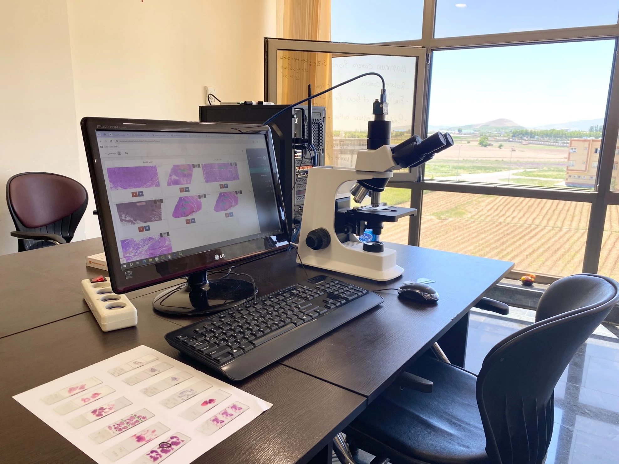
Digital pathology is the process of converting traditional glass microscope slides into digital images for easier analysis, management, and sharing. Pathologists can view and analyze high-resolution digital images on a computer screen instead of looking at physical slides under a microscope.
Here are some key aspects of digital pathology:
Digital pathology is revolutionizing traditional pathology by providing more efficient workflows, increased accuracy, and better scalability for clinical and research applications.
- Streamlining laboratory workflows for greater efficiency.
- Access to samples anytime and anywhere.
- Displaying all sample details on a monitor.
- Sharing high-quality samples with other professionals.
- Consulting with non-academic centers.
- Creating a digital archive of samples.
Digitizing pathology slides involves a precise process of converting glass samples into digital images used for medical diagnosis and analysis. This process is typically carried out as follows:
This process is exactly what DigiPat software facilitates. By digitizing pathology slides and converting them into high-resolution images, DigiPat plays a key role in improving diagnostic accuracy and speed. DigiPat leverages digital pathology technology to provide more precise analysis, secure storage, and faster information sharing. This innovative technology not only reduces human errors but also modernizes traditional pathology systems, assisting doctors worldwide in accessing pathological data more efficiently.
Pathologists can quickly review slides without handling them physically. This accelerates the diagnostic process and reduces turnaround time.
Pathologists can access digital slides from anywhere in the world, making remote diagnosis and peer consultations easier. Digital pathology allows specialists in different regions to collaborate seamlessly, improving diagnostic accuracy, especially for rare or complex cases.
Eliminating the need for physical slide storage saves space and associated archive maintenance costs. Faster diagnoses and fewer errors also reduce operational expenses in laboratories and hospitals.
Slides can be digitally stored, eliminating physical storage needs and simplifying patient record and image retrieval.
It is completely natural for pathologists to think that digital pathology is extra work and more of a luxury than a necessity. Initially, they may believe that traditional microscopes are sufficient. Change is always challenging... However, once you gain a proper understanding of digital pathology and experience using DigiPat, you'll see how much faster and easier everything becomes. With DigiPat, you'll quickly realize that traditional methods cannot compete with digital solutions!
Since the beginning of 2024, the number of laboratories and hospitals equipped with digital pathology systems globally has been rapidly increasing, driven by technological advancements, AI integration, and the demand for faster and more efficient diagnostics. However, due to varying adoption stages in different regions, determining an exact count is challenging. In advanced markets like the United States, Europe, and Japan, digital pathology adoption is widespread, with institutions like Mayo Clinic leading the way in digitizing their pathology departments. In countries like Japan, entire educational and clinical systems are being adapted to digital methods from the outset. Globally, thousands of laboratories and hospitals have adopted digital pathology, although many are still in transition. Adoption rates vary by region, with North America and Europe leading, while Asia-Pacific and Latin America are catching up. The widespread shift towards digital pathology is supported by advancements in cloud computing and AI, which are expected to drive further growth.
© retko.ir. All Rights Reserved.
Designed by retko co